Understanding immunity at a cellular level

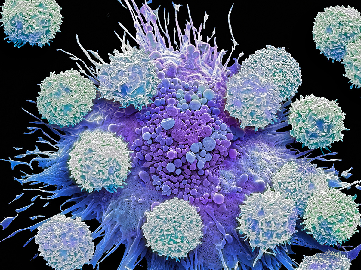
An immune cell called a macrophage. Research has found that lysosomes, an organelle that’s inside of such cells, expand in size when faced with infection.
New research into lysosomes, one of the specialized subunits inside cells called organelles, may help to further understanding of immune systems. Advancing knowledge of the function of lysosomes in the cellular immune system could help scientists to develop better vaccines or help to abate the impacts of autoimmune diseases.
Cell biology professor and Canada Research Chair in Organelle Function and Adaptation Roberto Botelho works with his lab team to deepen understanding of how cells achieve internal organization between the different functions of organelles.
Part of his research includes examining the role of lysosomes in immune cells called phagocytes when faced with infection. “They are specialized in that they roam our bodies, they hunt down infections, microbes, and then they go ahead and eat them. They literally engulf them,” he said of phagocytes.
How cells develop immunity
Lysosomes in these cells are essentially packets of acid and digestive enzymes – which professor Botelho describes as “basically little stomachs” – that digest the microbe by fusing with the phagosome that has surrounded the microbe. Depending on the type of immune cell, this fusion will either completely destroy the microbe or save some little pieces called antigens, substances that create an immune response, and put the remains on the outside of the cell.
Another type of immune cell, a T-cell, then examines these antigens and learns to recognize them as a foreign object that should be destroyed, teaching the body to make antibodies.
“This is how you get adaptive immunity,” said professor Botelho. This immunity can be developed through either having the disease or via vaccinations. Previous research has shown that when facing an infection, an immune cell’s lysosomes change shape from spheres to long tubes. But recent research found that in addition to the shape change, the lysosomal system actually grows rapidly and substantially, so that the immune cells have double the number of lysosomes. The process happens in approximately two hours. Professor Botelho and his lab team and collaborators, including PhD student Victoria Hipolito and professors Ivan Topisirovic of McGill University and Ola Larsson of Sweden’s Karolinska Institutet, published their research findings in Plos Biology.
“By expanding the lysosomal system, you now have twice the stomach, where [the lysosome] can eat more, so you can take more material and more antigen,” he said. This makes it more likely for the process of placing antigens on the surface of the cell for T-cells to analyze and recognize as foreign to succeed. “If you mess that up, cells do not present as well,” he said. “The whole process is less efficient.”
Professor Botelho and his collaborators are continuing research into the lysosomal system expansion by examining how the process works in a living organism. He said the process is at the core of immunity, and deepening understanding of how it works could lead to smarter, more efficient vaccines or the mitigation of autoimmune responses that make antibodies against the body’s own proteins.
Support for this research was provided by the Canadian Institutes of Health Research, the Canada Research Chairs program, the Government of Ontario, the Fonds de Recherche du Québec – Santé, the Wallenberg Academy Fellows program, the Swedish Research Council and the Swedish Cancer Society.
Related links:
How a Ryerson-led team is developing an ultra-sensitive testing technology for COVID-19 and beyond
A world-first: Assessing the quality of kidneys before transplantation using photoacoustic imaging
How yeast may hold answers for more effective cancer treatments